Recent publications have demonstrated significant improvement in IVF treatment outcomes by implementing aCGH or SNP-based preimplantation genetic screening (PGS) (Bisignano A. et. al., 2011, Harper J. et. al., 2010). A highly attractive possibility of correlation between embryo morphology and chromosomal status has been discussed by some authors (Finn A., et. al . 2010, Alfarawati S., et. al. 2011).
The objective of this study was to identify the relationship between blastocyst morphology and euploidy rates analyzed by SNP and aCGH-based PGS.
A retrospective study of PGS outcome data from blastocysts biopsied on day 5 or day 6 was conducted to identify differences in euploidy rates in embryos analyzed by SNP or aCGH-based PGS.
All embryos were hatched on day 3 and were biopsied on day 5 or on day 6. Only good and fair quality embryos that had at least 3-7 herniating cells and met criteria for cryopreservation were considered for biopsy. Poor quality blastocysts were biopsied only per MD or patient request.
A total of 1003 embryos were analyzed for euploidy rates and blastocyst morphology. Morphology of blastocysts was evaluated by Gardner classification (Gardner et al., 1999). Embryos were divided into two groups based on blastocyst expansion (expanded and not expanded) and into three groups based on blastocyst quality: good (AA/AB/BA), fair (BB), and poor (-C or C-). Euploidy rates were assessed in each study group by SNP and aCGH PGS.
Euploidy rate was significantly higher for embryos available for biopsy on day 5 than for embryos available for biopsy on day 6 – 66.9±4.7 percent and 52.6±3.9 percent, respectively, regardless of degree of blastocyst expansion (p<0.05). Average number of blastocysts biopsied per case was 5.8±3.3 (Fig. 1). Time when embryos become available for biopsy – on day 5 or on day 6 – reflected dynamics of embryo development in vitro.
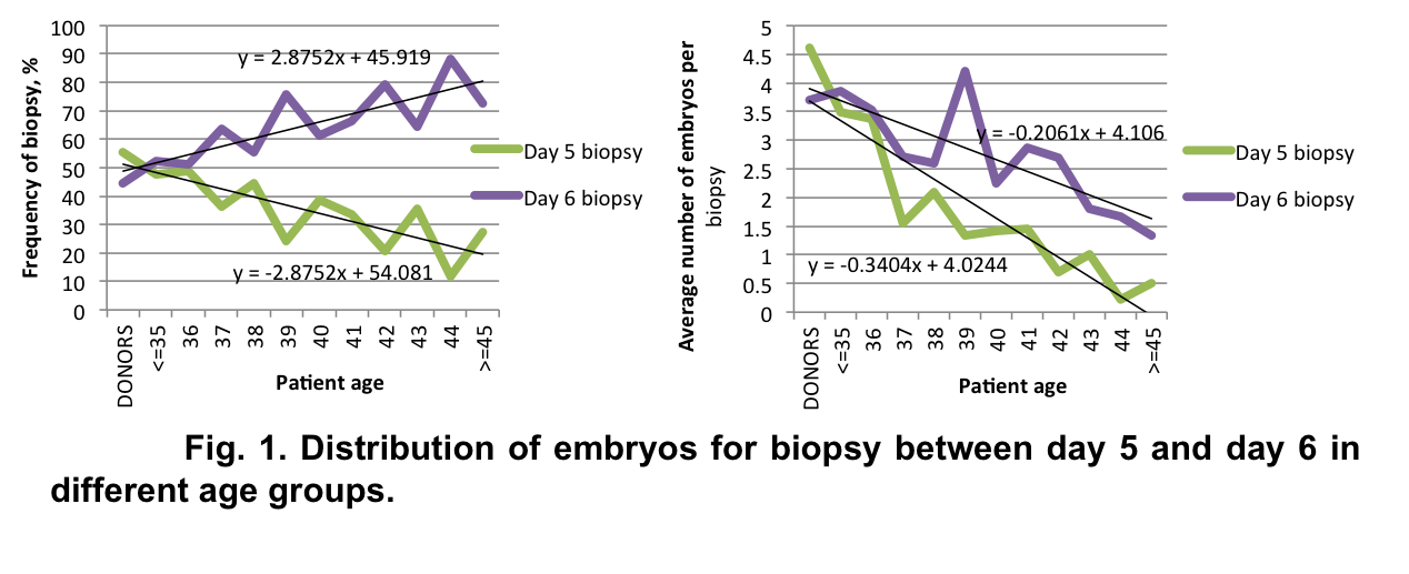
The difference in euploidy rates between embryos biopsied on day 5 vs day 6 had the tendency to increase with advancing patient age (Fig. 2).

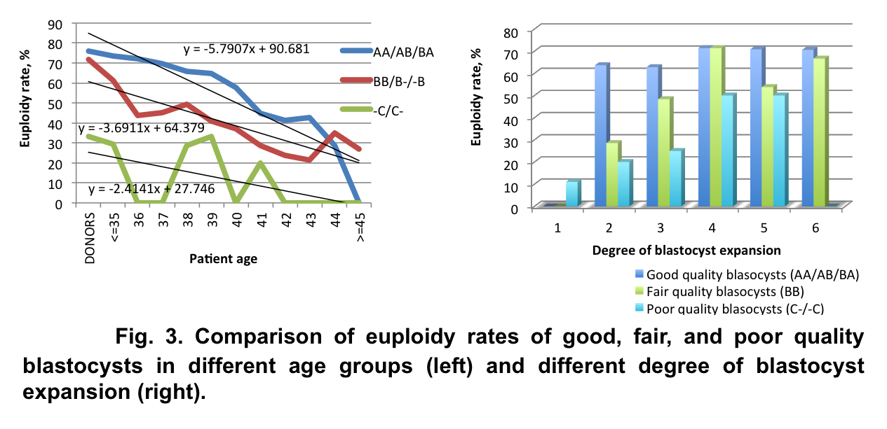
We observed a strong correlation between euploidy rates and embryo quality within the same patient age group: good quality blastocysts had a much higher rate of euploidy than poor quality blastocysts (Fig. 3).
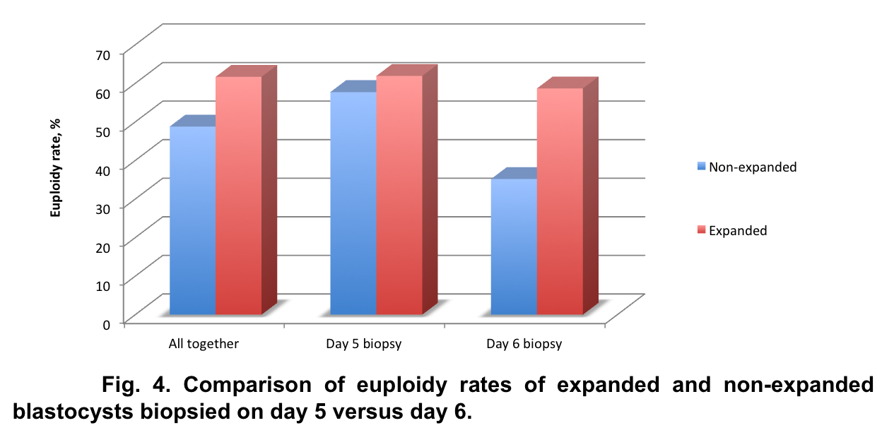
Our data demonstrated reliable differences in the number of euploid blastocysts on day 5 and day 6 among non-expanded blastocysts (57.7±7.1 percent and 35.2±4.9 percent, respectively, p<0.05). There was no statistically significant difference in euploidy rates in expanded blastocysts on day 5 and day 6 – 61.9±8.3 percent and 58.7±6.9 percent, respectively, p=0.4591 (Fig. 4).
Analysis of chromosomal status of blastocysts with different morphology demonstrated association of euploidy rates not only with quality of the embryos but also with degree of blastocyst expansion within the same quality group (Fig. 5).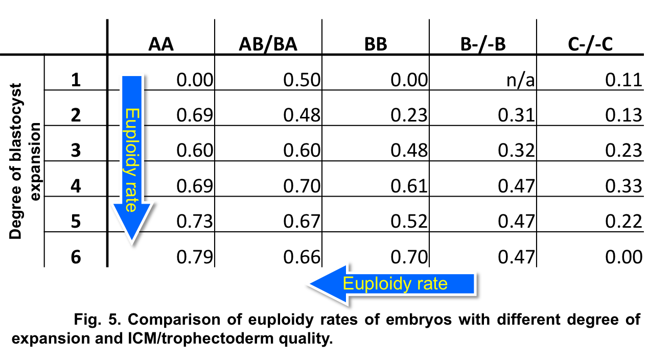
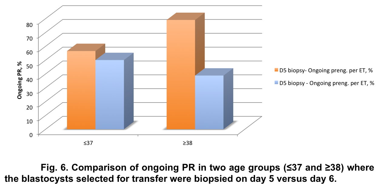
Our data demonstrated a statistically significant difference (p<0.05, χ2=20.2032) in pregnancy rates between cases where embryos selected for ET were biopsied on day 5 versus day 6. The difference in clinical pregnancy rate was even more dramatic for women over 38 years of age (mean age – 40.1±1.8 years): 79.0% for day 5 biopsy and 38.8% for day 6 biopsy (Fig. 6).
Overall euploidy rates defined by SNP were very similar to aCGH (52.4±8.1 percent and 55.1±6.1 percent, respectively, p=0.159707) and were not statistically different in all study groups.
Conclusion
Comprehensive chromosome screening provides an opportunity to re-evaluate correlation between embryo euploidy and embryo morphology in order to create the best treatment strategy for the infertile couple undergoing IVF treatment. There is a significant difference in euploidy rates depending on quality of the blastocysts and time they become available for biopsy; however, our data demonstrated no difference between the use of SNP and aCGH-based PGS to determine euploidy rates in human blastocysts.
References
2.Harper JC, Harton G. The use of arrays in PGD/PGS. Fertil Steril 2010; 94:1173 – 1177.